
Three canals on the radiograph, four in the tooth. What research says about root canal treatment failure — and what dental students can do about it before graduating.
You’re in the clinic during your internship. Upper first molar, hand files, a tutor somewhere across the room with another four students waiting for help.
The access cavity is open. You can see three orifices: mesiobuccal, distobuccal and palatal. The working-length radiograph looks reasonable. Everything resembles the diagram you studied, and the case feels as though it is going well.
Except there may be another canal.
In Indian populations, CBCT-based research suggests that an MB2 canal may be present in roughly two-thirds of maxillary first molars.
That gets close to one of the real problems behind root canal treatment failure. Sometimes treatment does not fail because the clinician handled a file badly. It fails because part of the anatomy was never found in the first place.
If you’re in second or third year and haven’t touched a molar yet, that’s worth knowing now. The habit of expecting three canals can form long before your first independent case.
How common is the MB2 canal in maxillary first molars?
A systematic review and meta-analysis of Indian populations looked at the prevalence of the second mesiobuccally canal — MB2 — in permanent maxillary first molars.
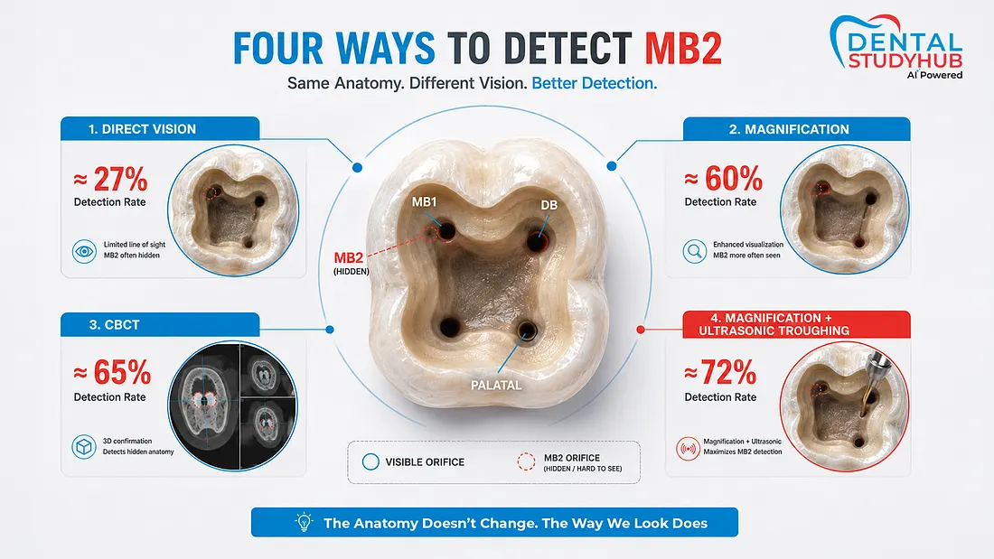
The interesting part isn’t just the prevalence. It’s how much the detection rate changed depending on how clinicians looked for it:
- Around 65% with CBCT
- Around 27% with direct vision
- Around 60% with magnification
- Nearly 72% with magnification plus ultrasonic troughing

Global pooled estimates run higher still, at roughly 70% for maxillary first molars and around 39% for second molars.
The anatomy didn’t change between 27% and 65%. Only the looking did.
That distinction matters in an undergraduate clinic.
You may be working without a microscope, with limited magnification, while your supervisor moves between several chairs. Sometimes the overhead light seems better positioned to illuminate your fingers than the floor of the pulp chamber.
It sounds like a small thing. It matters more than it should.
And missed anatomy is not harmless. A CBCT analysis involving mandibular second molars — a different tooth, but the same underlying problem — reported a high frequency of treatment failure when canals had been missed.
Another study of 772 endodontically treated teeth found missed canals in 13.3% of cases, with maxillary first molars accounting for a large proportion of the untreated canals identified.
The useful lesson for a student is simpler than the statistics: before you can find unusual anatomy, you have to expect it to exist.
There isn’t a satisfying shortcut for practising this when your college has no microscope and the shared apex locator is, once again, out of charge. Loupes help. Extracted teeth help, if your department gives you access to them.
But expectation costs nothing.
Know where the extra canal is likely to be before you start looking.

So what actually causes root canal failure?
At a biological level, root canal treatment usually fails because infection persists or returns. Clinically, that can happen because anatomy was missed, working length was inadequate, areas of the canal remained untouched or insufficiently disinfected, or the coronal seal later allowed reinfection.
In a textbook, these problems appear in different chapters.
Inside a tooth, they become one continuous problem.

Missed anatomy: what you never found
A canal that is never located cannot be shaped, irrigated or obturated.
That sounds almost too obvious to say. It becomes less obvious when you’re staring at a pulp chamber that appears to contain exactly the number of orifices your undergraduate diagram told you to expect.
MB2 is the familiar example, but the larger lesson is about anatomical variation.
Root canal anatomy is not obliged to resemble the illustration you memorised.
And finding the canal is only the beginning.
Working length: where you stop matters
Working-length errors matter in both directions. Root fillings ending substantially short of the desired apical endpoint have been associated in some studies with poorer outcomes, while overextension beyond the apex creates a different set of problems.
A systematic review of randomised trials reported acceptable working lengths in approximately 87–92% of cases using electronic apex locators, compared with around 74–83% using radiographic methods.
But the evidence isn’t completely one-sided.
An umbrella review found a more complicated picture. Some analyses showed comparable strict accuracy, while apex locators performed better on measures of clinical adequacy.
That gives you a much more useful answer for a viva than simply saying one method is “better.”
The apex locator helps estimate the apical endpoint electrically. The radiograph gives you morphology, curvature and surrounding anatomy.

Use them together. They answer different questions.
What the file never touches
This is where the phrase cleaning and shaping becomes slightly misleading.
A file does not contact every part of the root canal system.
Micro-CT research has shown substantial areas of canal wall remaining untouched after mechanical preparation. In curved mesial canals of mandibular molars, reported figures have ranged from roughly 20% to 42% under different experimental conditions. Some distal-root studies have reported still higher proportions.
Different rotary and reciprocating systems shift those numbers.
None makes the anatomical problem disappear.

Which changes how you should think about shaping.
The file is not cleaning every surface of the canal. It is creating the conditions that allow disinfection to reach places the instrument cannot.
Why irrigation matters so much
Once you accept that instruments leave parts of the canal untouched, irrigation stops looking like the liquid step performed between files.
It becomes part of the biological treatment.

Research comparing irrigation approaches suggests that activation can improve biofilm disruption. For example, research on ultrasonic activation of sodium hypochlorite reported significantly greater bacterial biofilm reduction than sonic activation or conventional needle irrigation in the models studied.
The important word there is reduction, not sterilisation.
Neither an instrument nor an irrigate gives you a perfectly sterile canal system.
Sequence may matter as well, with evidence favouring sodium hypochlorite followed by EDTA over the reverse sequence under the conditions studied.
Concentration, contact time, volume, activation and needle placement all contribute. Each decision may seem minor in isolation, but across several canals in a molar, those decisions compound.
And then there is the coronal seal
Students naturally spend enormous attention on the root filling. That’s understandable: it is visible on the final radiograph and easy to judge.
But treatment does not end when the gutta-percha reaches the correct length.
Ray and Trope’s well-known assessment of 1,010 endodontically treated teeth, published in the International Endodontic Journal in 1995, reported a strong relationship between the quality of the coronal restoration and periapical status.
Later research has supported parts of that relationship while making the overall picture more nuanced.
The useful conclusion isn’t that restoration matters and obturation doesn’t.
It’s that good endodontics can still be undermined by a compromised coronal seal.
Better materials do not remove the need to get the basics right either. A 2025 meta-analysis of randomised trials examining single-cone bio ceramic obturation reported high success rates, but differences compared with control techniques did not reach statistical significance at the reported follow-up intervals.
The material is rarely the whole story.
No sealer can disinfect anatomy that was never located.
Endodontic or periodontal? Sometimes the harder problem comes before treatment
Endo-perio lesions can punish diagnostic errors more reliably than technical ones.
A tooth with a deep isolated periodontal pocket and an equivocal pulp response could represent very different biological situations: a primary endodontic lesion draining through the periodontal tissues, periodontal disease with secondary pulpal involvement, combined endodontic-periodontal disease, or root damage such as a vertical root fracture.
Those are not interchangeable diagnoses, and they do not carry the same prognosis.
The 2018 World Workshop classification helped organise endodontic-periodontal lesions according to factors including whether root damage is present. But real patients remain messier than classifications.
Pulp sensibility testing depends partly on the patient’s response. Patterns of bone loss can overlap. Radiographs flatten three-dimensional anatomy into two dimensions.
Research comparing periapical radiographs alone with radiographs plus CBCT found improved diagnostic accuracy when CBCT information was added in the endodontic-periodontal cases studied.
But the scan doesn’t generate the hypothesis.
You still need to know what you’re trying to distinguish.
For a dental student, that’s the larger lesson: diagnostic tools become more useful when they are answering a clinical question you have already formed.
History, pulp testing, periodontal findings and imaging work better together than they do as isolated boxes in an examination.
What should a BDS student actually remember from this?
Probably not every percentage.
Three ideas are more useful.
Expect variation before you look for it
If you assume every maxillary first molar contains the anatomy shown in a basic textbook diagram, additional anatomy becomes easier to miss.
Expectation changes how carefully you inspect the chamber.
Think of shaping as preparation for disinfection
Instrumentation is essential, but instruments cannot contact every part of a complex root canal system.
The shape you create has a biological purpose: it gives your irrigation and disinfection strategy better access.
Stop looking for one perfect diagnostic tool
Apex locator or radiograph? Pulp test or CBCT?
Those questions sound clean in an exam.
Real diagnosis usually works better when different tools answer different parts of the problem. Clinical reasoning is often about combining imperfect information.
That is harder to teach than a file sequence.
It is also more transferable.
Where an endodontics course fits — and where it doesn’t
Most short endodontic programmes advertised to dental students and young dentists are understandably hands-on.
Rotary instrumentation, torque settings, file sequences, obturation.
Those courses can be useful.
But they work best when you already understand what you’re trying to achieve biologically and diagnostically.
If you’re still uncertain about canal morphology, working length, irrigation chemistry, endodontic microbiology or the endo-perio differential, learning another file sequence does not automatically close that gap.
Skill training works better when there is already a conceptual framework to attach the skill to.
That is why we built the Dental Study Hub Endodontics course as the step before advanced hands-on training — not as a replacement for it.
The online Endodontics course for dental students at Dental Study Hub runs across nine modules covering:
Pulpal and peri radicular pathology & diagnostic aids · Endodontic instruments · Working-length determination · Shaping and cleaning · Irritants, sealers & medicaments · Obturation · Endodontic microbiology · Endo-perio relationships · Endodontic microsurgery & lasers
It runs for three months, is self-paced, costs ₹8,000, and includes a certificate that can be added to your professional profile.
There is, however, an important limitation.
It won’t teach your hands anything. No online course can.
If your diagnostic and biological foundation is already strong and your main gap is psychomotor skill, this probably isn’t where you should spend your money. Put it towards supervised hands-on training.
If, however, you want to understand why a canal is searched for, why working length matters, why irrigation can change the biological outcome, and why two apparently similar radiographs may represent very different diagnoses, structured theory can make limited clinical exposure considerably more useful.
The aim is not to replace the molars you need to treat.
It is to make the molars you do get during BDS teach you more.
A few questions students usually ask
Why do root canals fail most often?
Persistent or recurrent intracanal infection is the central biological reason many root canal treatments fail. Contributing factors can include missed anatomy, incomplete cleaning and disinfection, inadequate working length, persistent microorganisms and a compromised coronal seal.
Can a missed MB2 canal cause root canal failure?
Yes. An untreated canal can retain infected or necrotic tissue and microorganisms that were never exposed to the intended cleaning, disinfection and obturation procedures. MB2 matters particularly because it is common in maxillary first molars and can be difficult to detect clinically.
How is the MB2 canal located without a microscope?
Expectation comes first. In a maxillary first molar, the operator should be aware that a second mesiobuccally canal is common and carefully inspect the developmental anatomy between the main mesiobuccally and palatal regions. Magnification and appropriate access refinement can improve detection. Clinical techniques should always be learned and performed under appropriate supervision.
Is an apex locator more accurate than a radiograph?
Evidence from randomised trials generally favours electronic apex locators for obtaining clinically acceptable working-length estimates, but radiographs provide morphological information an apex locator cannot. In practice, the two are complementary rather than interchangeable.
Why doesn’t instrumentation clean the entire root canal?
Root canal systems contain fins, recesses, isthmuses, curvatures and irregular surfaces that files cannot mechanically contact. Micro-CT studies consistently show untouched canal-wall areas after instrumentation, which is why irrigation and disinfection remain essential.
Is an online endodontics course worth it for BDS students?
For diagnostic principles, biological foundations, anatomy, working-length concepts, irrigation and endodontic reasoning, online study can be useful. For tactile and psychomotor skills, it cannot replace supervised hands-on practice.
A sensible sequence is:
Understand the reasoning → practise under supervision → refine the technique.
Back to that molar
Three orifices. Three canals on the radiograph. And reasonable odds there was another canal somewhere in the tooth.
You’ll treat more molars after graduation. Some will be straightforward. Some will remind you very quickly that teeth have never read your textbook.
The important question is whether anything changes between the first difficult case and the next hundred.
The file didn’t fail. The looking did.
And looking is a knowledge problem long before it becomes a technical one.
Which is why it is something you can start improving while you’re still a student.
Start with the free trial of the Dental Study Hub Endodontics course. Explore the pathology and diagnostic content first, then decide whether the full course fills the gap you actually have. No payment upfront.
References
Evidence-Based Dentistry (2022) — systematic review and meta-analysis of MB2 prevalence in Indian populations
International Endodontic Journal — global pooled MB2 prevalence, PMID 31735252
Journal of Clinical Medicine (2024);13(10):2931 — missed canals and treatment outcomes
PMC10478193 — prevalence of missed canals in endodontically treated teeth
PMC12590451 — systematic review of electronic apex locator accuracy
PMC11510102 — umbrella review of working-length determination
BMC Oral Health (2025) — micro-CT assessment of untouched canal walls
PMC11508828 — irrigation activation and bacterial biofilm reduction
PMC12563735 — irrigant sequence
Ray HA, Trope M. International Endodontic Journal. 1995;28:12–18
PMID 12196221 — coronal restoration quality and periapical health
Journal of Oral Biology and Craniofacial Research (2025) — bioceramic obturation meta-analysis
PMC12872107 — CBCT and endodontic-periodontal diagnostic accuracy
This article is for educational purposes and does not replace formal clinical training, supervision or case-specific professional judgement. Endodontic outcomes vary across populations, operators, case selection, follow-up periods and definitions of treatment success; numerical values are reported in the context of the cited studies.


Comments (0)
No comments yet. Be the first to share your thoughts!